An HRCT chest scan can reveal fine changes in lung tissue, small airways and surrounding chest structures, but the exact images depend on the protocol ordered. By the end, you will know what a plain HRCT includes, how it differs from other chest scans, how to prepare and which booking details to confirm.
Key takeaways
- HRCT shows lung tissue, small airways, pleura and nearby chest structures.
- HRCT helps assess interstitial lung disease, fibrosis, bronchiectasis and air trapping.
- HRCT chest plain means no contrast dye was used for the scan.
- Confirm the protocol, scan range, contrast requirement and report turnaround before booking.
Which parts of the chest can an HRCT scan show?
Thin-section HRCT images show the lung tissue in greater detail than a routine chest X-ray. The main target is the pulmonary interstitium and small airways, but the report also covers the pleura, mediastinum, heart, chest wall and visible upper abdominal structures. The exact hrct scan inclusions depend on the scan range and protocol.
| Region | What HRCT can show | Patterns doctors assess |
|---|---|---|
| Lung tissue | Interstitium, alveoli and blood-vessel markings | Ground-glass opacity, reticulation, interlobular septal thickening and honeycombing |
| Airways | Bronchi and smaller airways | Bronchiectasis, airway-wall thickening, mucus plugging and air trapping |
| Pleura | The lining around each lung and pleural space | Thickening, plaques, fluid, air or irregular pleural deposits |
| Mediastinum and hila | Central chest structures, lymph nodes and major vessels | Enlarged lymph nodes, masses, vessel enlargement or compression |
| Chest wall and upper abdomen | Ribs, muscles, diaphragm and the uppermost liver, spleen or stomach | Fractures, soft-tissue changes or incidental abnormalities |
Images taken during full inspiration show how much lung fills with air. Expiratory images can reveal air trapping from small-airway disease, while prone images can separate gravity-related dependent atelectasis from genuine posterior lung abnormality. These additional series are not automatic in every HRCT.
A pattern such as ground-glass opacity or fibrosis can narrow the diagnostic possibilities, but it does not identify the cause alone. The scan also cannot measure breathing capacity; pulmonary-function testing and clinical assessment may still be needed.
What conditions can HRCT help evaluate, and what are its limits?
HRCT can investigate diffuse or patchy lung abnormalities, especially when a chest X-ray is abnormal, symptoms persist, or the suspected disease affects small airways or the pulmonary interstitium. It can reveal ground-glass opacity, reticulation, interlobular septal thickening, honeycombing, bronchiectasis, emphysema, air trapping and small nodules.
- Interstitial lung diseases, including patterns associated with pulmonary fibrosis
- Bronchiectasis and chronic airway damage
- Emphysema and smoking-related lung disease
- Small-airway disease, including bronchiolitis with air trapping
- Infection, including persistent or complicated infection
- Pulmonary nodules and unexplained shadows or opacities seen on an X-ray
An HRCT pattern narrows the possibilities; it does not always identify the disease. Ground-glass opacity, for example, can result from infection, inflammation, fluid, haemorrhage or early fibrosis. The radiologist needs your symptom duration, smoking or occupational exposure, immune status, previous infection and earlier scans before assigning significance.
HRCT also shows structure, not performance. A scan can reveal emphysema or fibrosis without measuring how much air you move, how well oxygen crosses into blood, or how exercise affects you. Spirometry, lung volumes, diffusion capacity and oxygen assessment answer those functional questions.
A stable-looking scan can coexist with impaired function, while a striking abnormality may have little effect on breathing. The report’s impression and clinical correlation matter more than one isolated image.
What does “HRCT chest plain” mean, and when is another scan better?
“HRCT chest plain” means a high-resolution CT of the chest performed without injected iodinated contrast. It uses thin sections, often about 1–1.5 mm, to show lung tissue, interstitial markings and small airways in detail. A plain HRCT is not a CT pulmonary angiogram (CTPA), and a normal result cannot reliably exclude a pulmonary embolism.
| Examination | What it adds | When it is the better choice |
|---|---|---|
| Plain HRCT | Thin-section detail of lung tissue and small airways without contrast | Suspected interstitial lung disease, fibrosis, bronchiectasis or emphysema |
| Contrast-enhanced chest CT | Highlights vessels, pleura, mediastinum and selected masses | A mass, pleural problem, vascular question or mediastinal abnormality needs assessment |
| CTPA | Timed intravenous contrast outlines the pulmonary arteries | Pulmonary embolism is suspected; plain HRCT is not a substitute |
| Chest X-ray | Fast, lower-dose overview of the chest | Initial assessment, routine follow-up or checking a large change |
| MRI | Excellent soft-tissue detail without ionising radiation | Selected heart, chest-wall or mediastinal problems; it is not the usual test for fine lung detail |
| Pulmonary-function testing | Measures airflow, lung volumes and gas transfer | Breathlessness needs a functional assessment; CT images alone cannot measure lung performance |
Before booking an hrct chest plain scan in fursungi, confirm that the referral requests thin-section HRCT, states “non-contrast,” and specifies inspiratory or expiratory images when small-airway disease is suspected. Contrast plans can change preparation, so tell the centre about pregnancy, kidney disease and previous contrast reactions.
What does the HRCT appointment include, and how should you prepare?
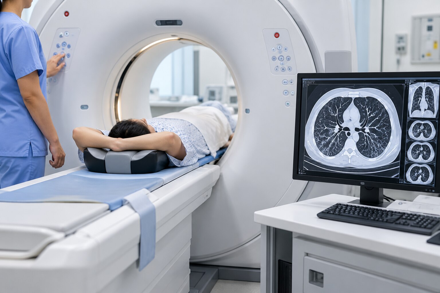
Your appointment usually covers the chest from the lung apices to the costophrenic angles, with thin axial sections of about 1–1.5 mm. The radiologist may add expiratory images to reveal air trapping or prone images to separate dependent atelectasis from genuine posterior lung disease; neither series is automatic.
1. Bring earlier CT images and reports. Comparison can prevent an unnecessary repeat scan and helps identify whether a finding is new, stable or changing.
2. For a plain HRCT, follow the centre’s usual chest CT scan preparation instructions; injected contrast is not used, and fasting is often unnecessary. Wear comfortable clothing, remove metal near the chest, and tell staff if pregnancy is possible.
3. For a contrast study, ask whether fasting is required and report previous iodinated-contrast reactions, kidney disease, diabetes, thyroid disease or pregnancy. The facility may request a recent creatinine or eGFR result before injection.
4. During the scan, you lie on a motorised table that moves through the gantry. Follow the breathing command and hold your breath when instructed; movement or an incomplete breath-hold can blur the lungs.
The study is reviewed in lung and mediastinal windows. Coronal and sagittal reconstructions supplement the axial images, helping show the height, length and distribution of abnormalities. You should receive a written radiologist’s report, plus access to the images through a disc, download link or viewing portal.
What should you confirm when booking an HRCT in Fursungi or Pune?
Before booking, confirm the exact protocol rather than relying on the word “HRCT.” This matters when comparing HRCT chest scan details in Fursungi with an HRCT chest scan Pune appointment.
1. Ask whether the scan uses thin sections, usually about 1–1.5 mm, with lung and mediastinal reconstructions. Confirm the scan range, from the lung apices through the costophrenic angles.
2. Confirm whether it is HRCT chest plain, meaning no injected iodinated contrast. Contrast is a different choice for suspected pulmonary embolism, vascular disease, a mediastinal or pleural problem, or mass characterisation.
3. Ask which image series the radiologist has requested: full-inspiration images, expiratory images for air trapping, and prone images when dependent atelectasis could mimic lung disease. These are not automatic in every examination.
4. Ask how you will receive the images: DICOM files through a portal, a disc or USB drive, and access to the written radiologist’s report. Confirm when the report will be available and keep earlier scans for comparison.
5. Tell the centre about pregnancy, previous CTs, symptoms, smoking or occupational exposure, and the doctor’s specific question. That information helps the radiologist interpret an otherwise nonspecific pattern.
When arranging the scan at Shivam Hospital M-08042754125 in Fursungi, ask these questions before fixing the appointment so the booked examination matches the referring doctor’s request.
Related product
Radiology HRCT Chest Plain Scan ₹4000 High-resolution computed tomography (HRCT) is a type of computed tomography (CT) with specific techniques to enhance image resolution. |
Frequently asked questions
Which parts of the chest can an HRCT scan show?
Thin-section HRCT images primarily show the lung interstitium and small airways. The report can also assess the pleura, mediastinum, heart, chest wall and visible upper abdominal structures, depending on the scan range.
What conditions can HRCT help evaluate?
HRCT can help evaluate interstitial lung disease, pulmonary fibrosis, bronchiectasis, emphysema, small-airway disease, air trapping and selected infections. It does not replace clinical assessment, pulmonary function tests or other scans when those are needed.
What does HRCT chest plain mean?
HRCT chest plain means the scan was performed without intravenous contrast dye. It is commonly used when detailed assessment of lung tissue and airways is the main goal; contrast CT may be better for blood vessels, some masses or specific complications.
How should you prepare for an HRCT chest scan?
Follow the imaging centre’s instructions about fasting, medicines and contrast. Wear comfortable clothing, remove metal objects, bring previous scans and reports, and tell staff if you are pregnant or could be pregnant.
What should you confirm when booking an HRCT in Fursungi or Pune?
Confirm that the centre offers the requested HRCT protocol, ask whether it is plain or contrast-enhanced, check the scan range, preparation instructions, report timing and whether you need to bring a prescription or previous images.
Keywords